When an egg is fertilized, it begins a remarkable journey. Over the next five days, it develops through several important stages before reaching the blastocyst stage, when it may be considered for transfer. During these five days, every developmental event matters.
Traditionally, embryologists have monitored embryos by taking them out of the incubator at specific intervals and observing them under a microscope. While this allows us to assess embryo development, every time an embryo is removed from its controlled environment, it is exposed to changes in temperature, gas conditions and light. Time-lapse technology offers a different approach. Instead of repeatedly taking embryos out for observation, it allows their development to be monitored continuously while they remain inside the incubator. In simple terms, it gives the embryologist a window into the embryo’s development without disturbing the culture environment. So, how does it work, and why is it becoming increasingly important in modern IVF laboratories?
Following fertilization through IVF or ICSI, the embryos are placed in controlled culture conditions where they develop over the next few days. During this period, embryologists monitor several important developmental events.These include the appearance and fading of the pronuclei (PN), the first cleavage, and subsequent divisions from 2 cells to 4 cells and then 8 cells. The embryo then progresses through compaction and forms a morula, followed by the blastocyst stage and, eventually, hatching. Each of these stages provides valuable information about embryo development. However, not every event happens neatly between two scheduled observations. Some developmental behaviours can occur quickly and may easily be missed during conventional manual assessment.
Incubators are at the heart of an IVF laboratory because they provide the environment embryos need to develop. Temperature, gas concentrations and humidity all need to be carefully controlled. Earlier incubators were designed to accommodate a large number of culture dishes. Although they provided a controlled environment, opening the incubator repeatedly could result in changes in temperature and gas conditions. The development of benchtop incubators improved this situation. Their smaller chambers allow environmental conditions to recover more quickly after opening. The next major development was the integration of imaging systems into incubators. This gave embryologists the ability to monitor embryo development continuously without repeatedly removing the embryos from their controlled environment. This is the basis of time-lapse embryo imaging.
One of the biggest advantages of time-lapse technology is the amount of information it can provide about embryo development. With conventional observation, an embryologist sees the embryo at particular points in time. With time-lapse imaging, the entire developmental journey can be reviewed. This can reveal events that may otherwise be missed during periodic observation. Events such as abnormal or trichotomous cleavage, reverse cleavage and other unusual developmental patterns can provide additional information when assessing embryos. Similarly, details such as blastocyst movement, compaction and incomplete compaction can be difficult to appreciate from isolated observations. This does not mean that time-lapse technology replaces the expertise of the embryologist. Rather, it gives the embryologist more information to work with when assessing embryo development.
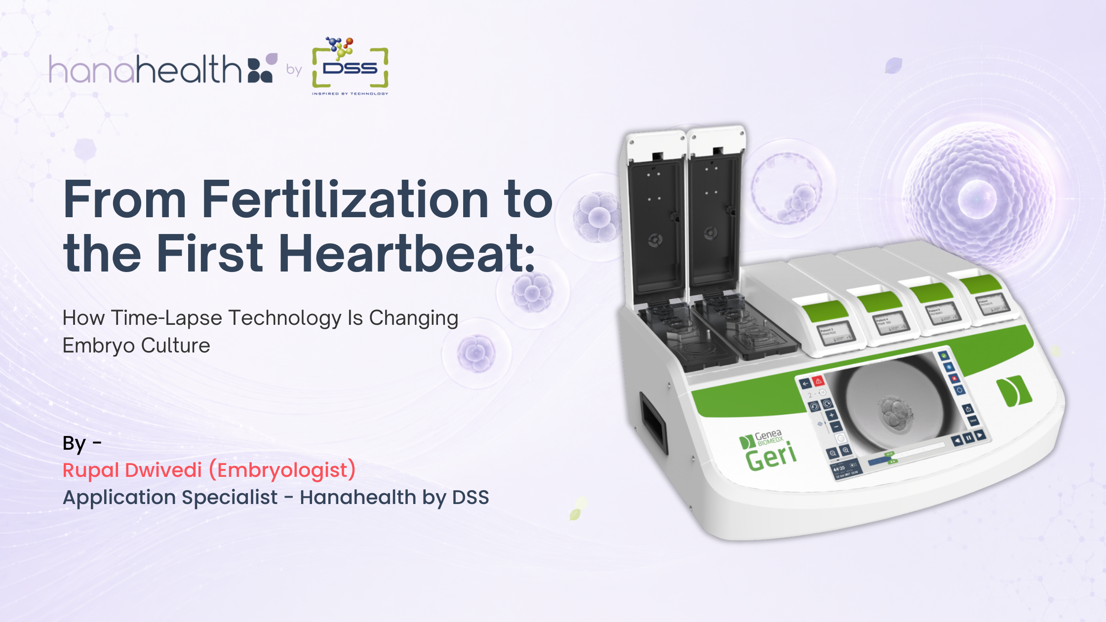
A time-lapse incubator can be described as the “eyes” of the embryologist inside the incubator. But not all systems are designed in the same way. A time-lapse system needs to do more than capture images. It must also provide a stable and reliable environment in which embryos can grow. This is where the GERI Time-Lapse Incubator by Genea Biomedx takes a different approach. GERI combines embryo culture and continuous imaging in a system designed around individual chambers. It has six independent chambers, each with its own dedicated imaging system. This means that each chamber can continuously monitor the embryos placed inside it without relying on a single camera moving between multiple positions. The system captures images every five minutes across 11 focal planes, allowing the development of the embryo to be followed in detail as it progresses through different stages. The images are then combined to create a time-lapse sequence, giving the embryologist a continuous view of embryo development. The imaging system also adjusts focus as the embryo develops, helping to maintain clear images throughout the culture period. And perhaps one of the most convenient features is that these images and videos can be assessed remotely, allowing embryologists to review embryo development without physically being in front of the incubator.
One of the key features of GERI is its individual chamber design. Each chamber has its own controlled gas supply, temperature monitoring and CO₂ and humidity sensors. This allows each chamber to function as an independent culture environment. Why is this important? In a conventional multi-chamber system, a problem affecting the overall incubator can potentially affect multiple patients at the same time. With independently controlled chambers, an issue in one chamber does not automatically compromise the conditions in the others.It also makes maintenance more practical. If one chamber requires servicing or replacement, the remaining chambers can continue operating. For an IVF laboratory, where continuity and reliability are extremely important, this level of independence can be a significant advantage.
GERI does not simply record embryo images. It continuously monitors the conditions in which those embryos are being cultured. Embryo culture is highly sensitive to environmental changes. Even relatively small deviations in temperature or gas conditions can be important. GERI therefore continuously monitors parameters such as temperature, CO₂ and humidity and provides alerts when values move outside the defined limits. The system also records these changes and presents them graphically. Instead of simply knowing that an alarm occurred, the embryologist can review the environmental history and see how the parameter changed over time.In other words, continuous monitoring is not just about collecting more data. It is about making that data useful.
An IVF laboratory generates a huge amount of information every day. A good system should not make that information more complicated; it should make it easier to understand.GERI is designed with this in mind. Each chamber has its own display showing information such as the patient’s name and UHID, along with temperature, CO₂ and humidity levels. The main touchscreen interface allows the embryologist to access individual embryos, review time-lapse videos and watch them at different playback speeds. Importantly, this can be done directly through the system without having to open the incubator or depend on separate software for basic viewing. Images and videos can also be exported when required. The system can provide access to information such as alarm history, environmental graphs and patient reports, giving the embryologist a consolidated view of both embryo development and culture conditions.
The real value of time-lapse technology is not simply that we can see more images.It is that we can understand the developmental story of an embryo in greater detail. GERI brings together controlled embryo culture, continuous imaging, environmental monitoring and data management in one system. Its individual chambers allow each group of embryos to have its own controlled environment, while dedicated imaging provides continuous monitoring of embryo development. For the embryologist, this means fewer disturbances to the culture environment and access to much more information than can be obtained from occasional manual observations alone. Embryo culture is a process in which small changes can matter. Every developmental event, every environmental fluctuation and every decision made along the way contributes to the bigger picture. Time-lapse technology gives embryologists the opportunity to see that picture more completely.
At its core, GERI is not simply taking pictures of embryos. It is continuously observing, recording and organising information so that embryologists can make better-informed decisions for their patients. From fertilization to blastocyst formation and hatching, the embryo is constantly changing. With time-lapse technology, we no longer have to rely only on snapshots of that journey; instead we can watch the entire journey.

IVF has helped millions of couples achieve their dream of parenthood, but it’s important to understand that success rates can vary significantly from one case to another. Here are the main factors that influence IVF success.
Read More
The field of In Vitro Fertilization (IVF) has come a long way since the birth of the first IVF baby in 1978. Recent technological .
Read More
When undergoing IVF, many factors can influence your chances of success, and lifestyle choices play a significant role.
Read More